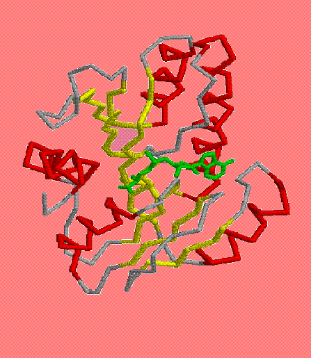
load H:\term1\credit_works\bespredel\1eiz.pdb
script H:\term1\credit_works\credit2\1eiz.def
echo This is my protein complexed with S-2adenosylmethionine

backbone 100 (1)
wireframe off
color green
select sam
wireframe 70
color magenta

    pause   (2)
    echo The nearest to the S-2Adenosylmethionine amino-acids
    select all
    backbone 20
    select sam
    wireframe 125
    select 64 or 65 or 99 or 124
    wireframe 100
    color cpk

        pause   (3)
        echo H-bonds between protein and S-2Adenosylmethionine
        restrict 64 or 65 or 99 or 124 or sam
        wireframe 50
        color cpk
        select sam
        wireframe 90
        color green
        select sam301.n or sam301.oxt or sam301.n6 or sam301.o
        select asp124.od2 or gly64.n or trp65.n or asp99.od1 | selected
        cpk 150
        zoom 200
        select sam301.n or sam301.n6 or gly64.n or trp65.n
        color blue
        select sam301.o or sam301.oxt
        color red
        center sam301.c1*
        bond 531 1413
        wireframe 15
        bond 723 1391
        wireframe 15
        bond 267 1394
        wireframe 15
        bond 263 1395
        wireframe 15

    pause   (4)
    echo stereoscopic picture of interaction between protein and S-2Adenosylmethionine
    restrict sam or cont_protein_SAM
    cpk 400
    select sam
    color magenta
    select cont_protein_SAM
    color greentint
    select sam301.n or sam301.n6 or gly64.n or trp65.n
    color blue
    select sam301.o or sam301.oxt or asp124.od2 or asp99.od1
    color red
(2)- показывает область взаимодействия белка с лигандом;
(3)- показывает само взаимодействие;
(4)- показывает объемное изображение взаимодействия белка с лигандом.