Standart protein as in most of exercises was used PDB ID 4ri1. Below is the comparison of the secondary structures found by stride -o 4ri1.pdb and described in the original pdb file. In both files residues are numbered the same making possible direct comparison. Protein of interest with coloring by ss is on the fig.1.
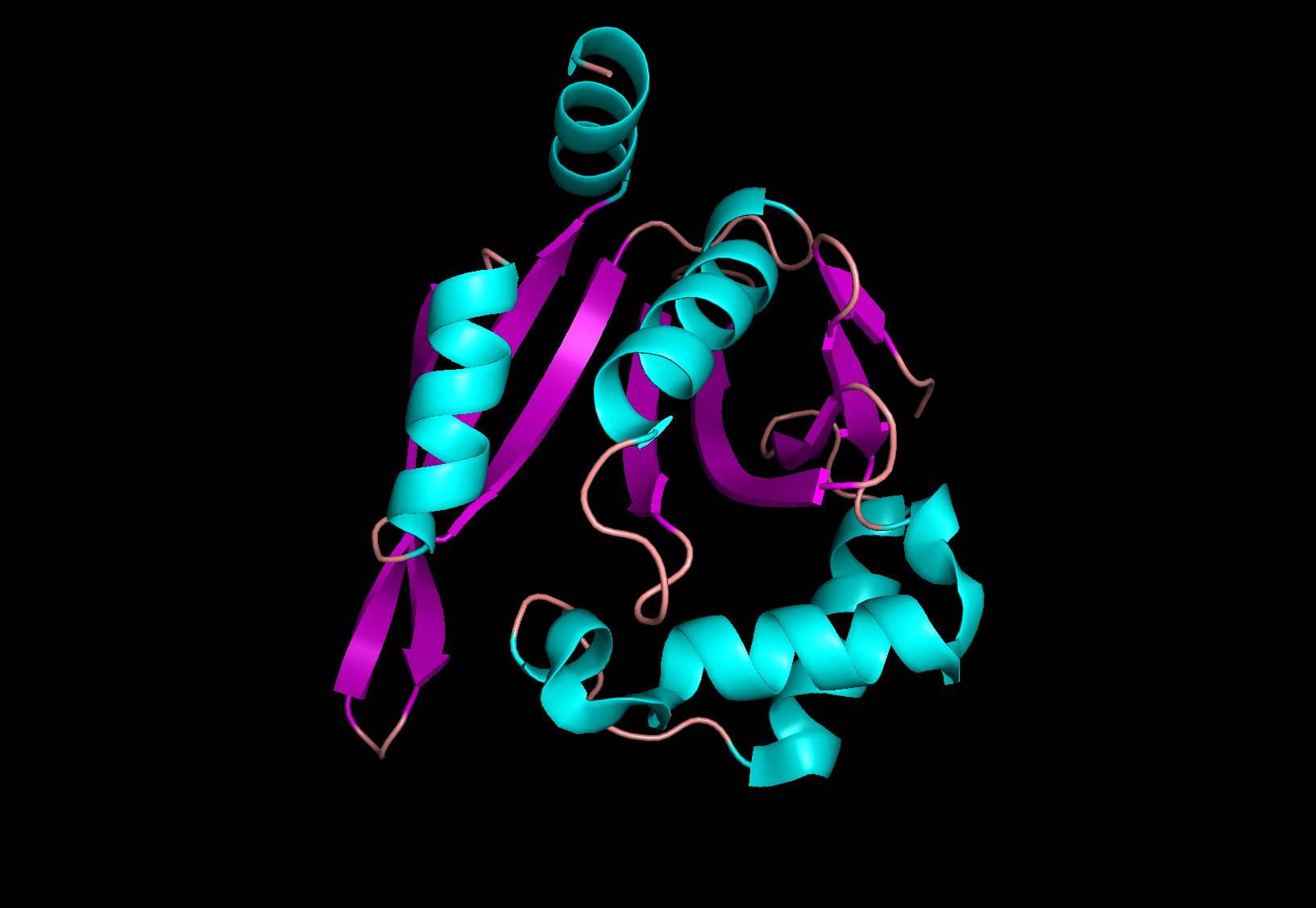
Figure 1. protein 4ri1 colored by ss
HELIX 5 5 GLY A     102    GLU A   116     1 15
HELIX 6 6 ASN A     131    ASN A   141      1 11
SHEET    1   A 8 TYR A   5  TYR A   7  0
SHEET    2   A 8 ILE A  10  ASP A  14 -1  O  ALA A  12   N  TYR A   5
SHEET    3   A 8 ARG A  63  GLU A  69 -1  O  LEU A  66   N  ILE A  13
SHEET    4   A 8 VAL A  72  ASN A  83 -1  O  GLY A  75   N  PHE A  67
SHEET    5   A 8 HIS A  88  LYS A  95 -1  O  TYR A  94   N  VAL A  76
SHEET    6   A 8 SER A 122  MET A 128  1  O  HIS A 124   N  GLY A  89
SHEET    7   A 8 GLU A 158  ASP A 168 -1  O  LEU A 163   N  VAL A 127
SHEET    8   A 8 GLU A 144  LYS A 155 -1  N  LYS A 155   O  GLU A 158
LOC  AlphaHelix   GLY   103 A      GLU    116 A                            4RI1
LOC  AlphaHelix   PHE   132 A      ASN    141 A                            4RI1
LOC  Strand       TYR     5 A      TYR      7 A                            4RI1
LOC  Strand       ILE    10 A      ASP     14 A                            4RI1
LOC  Strand       ARG    63 A      GLU     69 A                            4RI1
LOC  Strand       VAL    72 A      ASN     83 A                            4RI1
LOC  Strand       HIS    88 A      LYS     95 A                            4RI1
LOC  Strand       SER   122 A      MET    128 A                            4RI1
LOC  Strand       GLU   144 A      LYS    155 A                            4RI1
LOC  Strand       GLU   158 A      ASP    168 A                            4RI1
As it can be seen all beta-strands were predicted precisely by stride, and alpha-helixes were both off by one at the beginning.
Term 7 Main page