Data about unit cell from CRYST1 field of PDB file:
vector[a b c] = [107.830 145.620 166.190],
vector_angles[alpha beta gamma] = [90.00 90.00 90.00],
Spacegroup: P 21 21 21, this strands for trivial group with three axis of screw symmetry,
number of molecules in the cell = 24
Structure is composed of three chains shown on the fig. 1.
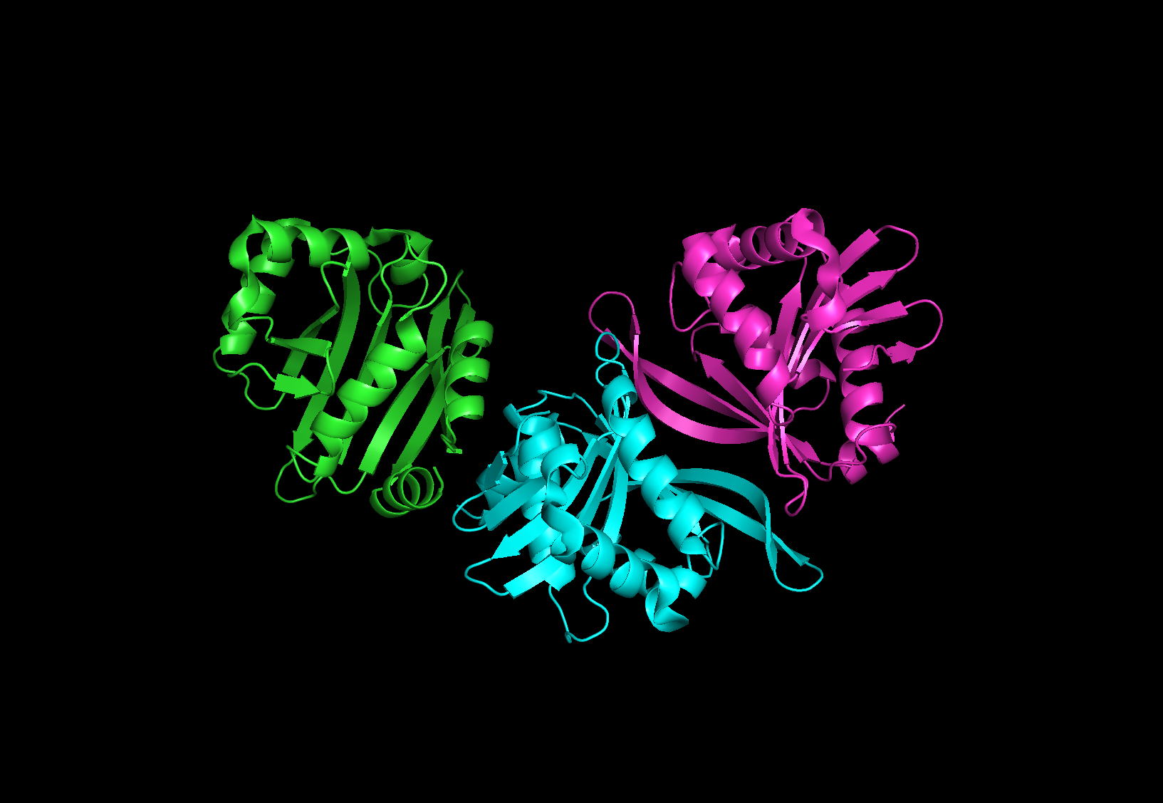
Figure 1, protein 4ri1
Structure of the protein can be seen on Fig.1. Three subunits compose
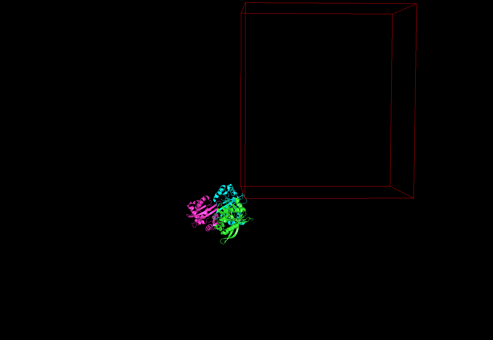
Figure 2, protein 4ri1 unit cell outlined
As can be seen in Fig.2 authors of the paper [1] did not position protein inside the unit cell, which is a bad practice.
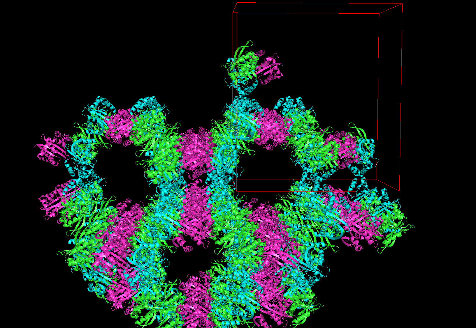
Figure 3, crystall lattice of 4ri1 protein
4ri1 protein is a dimeric in a natural form. And its assymetric unit in the lattice contains three chains.
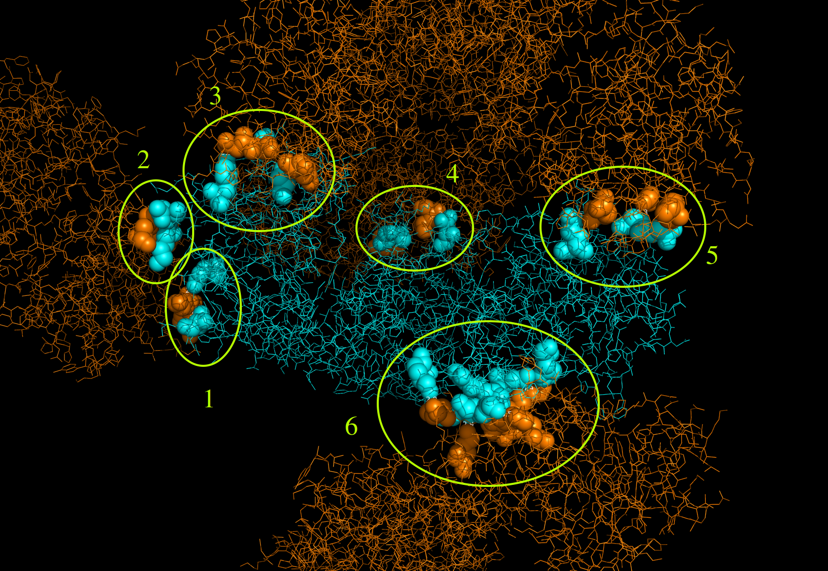
Figure 4, contact zones with proteins form other crystall cells
Six contact zones were discovered. Out of these six five have only couple of risidues connected with hydrogen bonds, thus they are highly unlikely to have biological significance and origin from artifacts of crystallization.
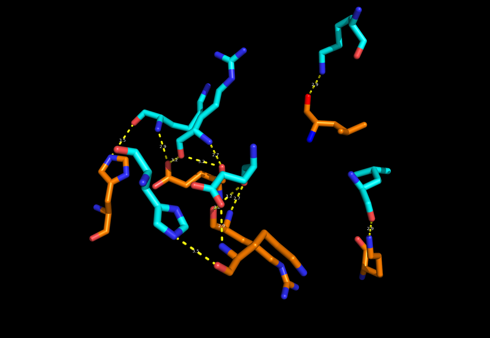
Figure 5, 6th contact zone
Closer look at the 6th contact zone shows enough hydrogen bonds to facilitate good binding and contact is very similar to that inside a native dimer (as also suggested in [1]). So it has biological relevance.
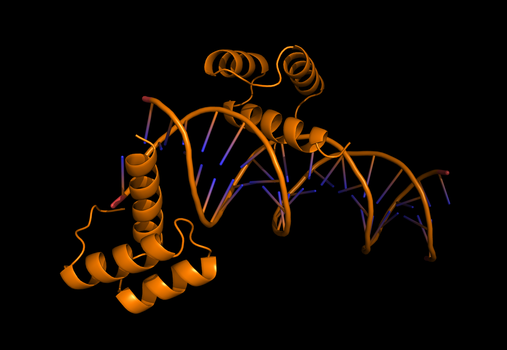
Figure 6, 3HDD complex
To demonstrate the necessity of adding adjacent symmetrycal unit to some DNA-protein complexes file with PDB ID: 3HDD was chosen (fig.6).
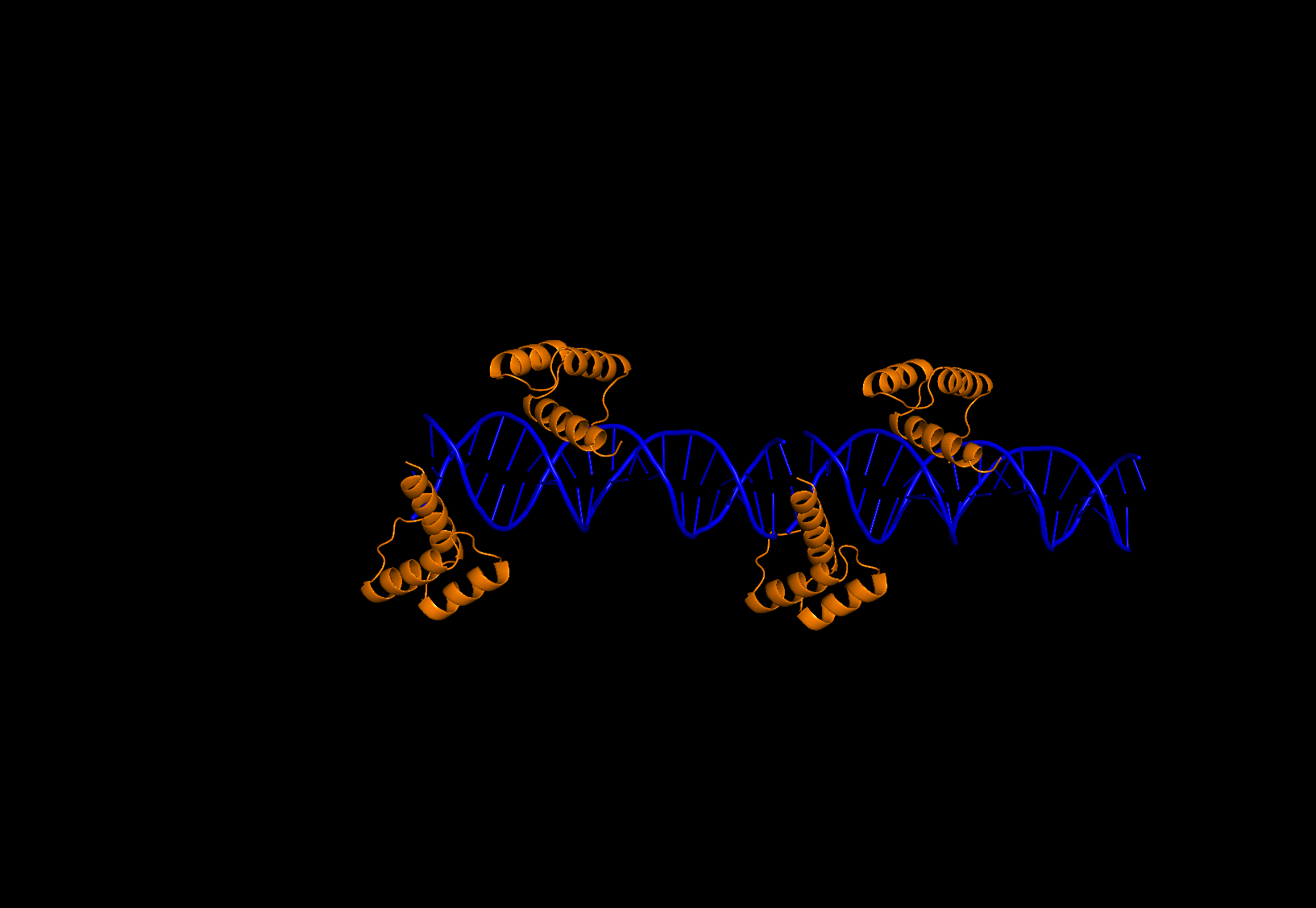
Figure 7, 3HDD complex with adjacent symmetrycal unit
Although on fig.6 it looks like the transcription factor is right on the edge of DNA, when complete picture on fig.7 is taken into account, one can clearly see that protein is interacting with double helix from another assymetric unit.
The asymmetric unit is the smallest portion of a crystal structure to which symmetry operations can be applied in order to generate the complete unit cell (the crystal repeating unit). Symmetry operations most common to crystals of biological macromolecules are rotations, translations and screw axes (combinations of rotation and translation).
The biological unit is the macromolecular assembly that has either been shown to be or is believed to be the functional form of the molecule. For example, the functional form of hemoglobin has four chains.
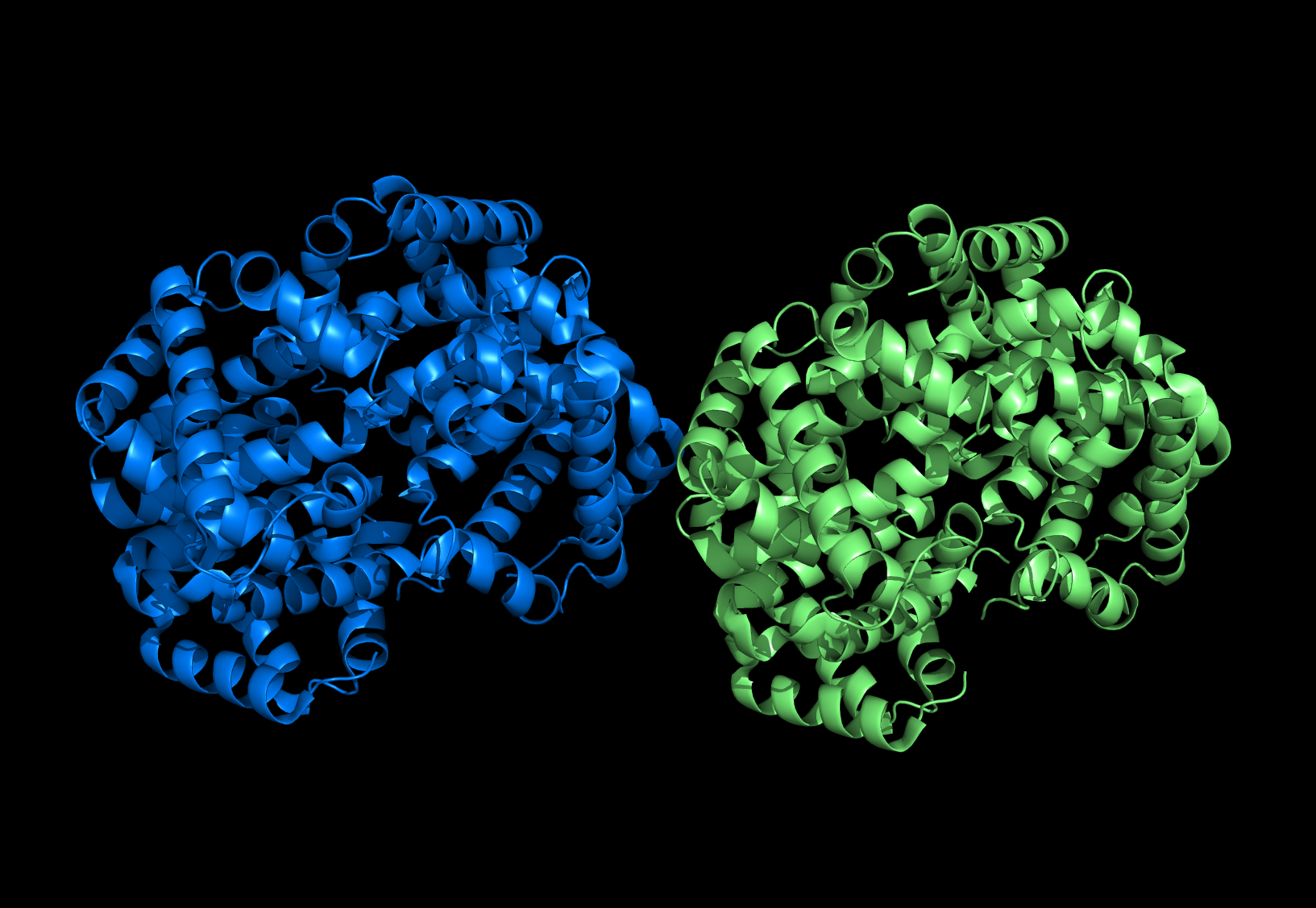
Figure 8, protein 1hv4
Protein PDB ID: 1hv4 is an example of difference between biological and asymmetric units its biological unit is only one half of asymmetric (shown in blue fig.8).
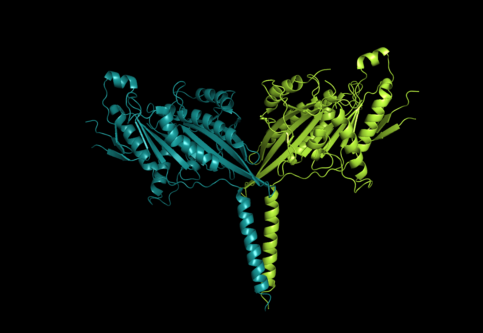
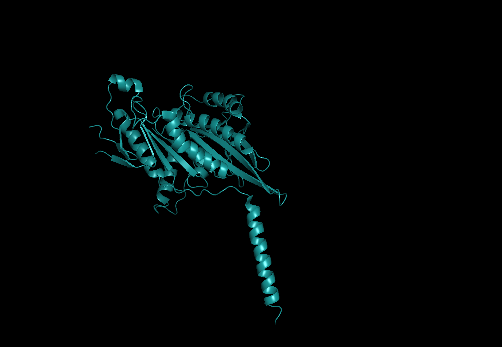
Figure 9,top - protein 6A1Z biological unit, bottom - assymetric
The opposite example is protein 6A1Z on fig.9 whose biological unit is a dimer of assymetric unit.
[1] Ud-Din, A.I., Liu, Y.C. and Roujeinikova, A., 2015. Crystal structure of Helicobacter pylori pseudaminic acid biosynthesis N-acetyltransferase PseH: implications for substrate specificity and catalysis. PLoS One, 10(3), p.e0115634.
Term 7 Main page