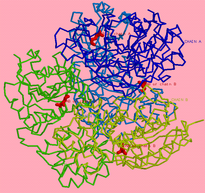
load H:\Term1\CreditWorks\Work\1djo.pdb
script H:\Term1\CreditWorks\Credit2\1djo.def
restrict none
background pinktint
restrict *a
backbone 100
wireframe off
restrict cab4001
wireframe 100
select within (2.0, *a) and cab4001
select within (4.5, cab4001) and *a
wireframe 30
select within (3.5, cab4001) and *a and not *a.c??
cpk 150
select cab4001
cpk off
zoom 600
center cab4001 and *.c2
select contact_mol
wireframe 30
restrict contact_mol, cab
select 4001 and *.c3
label %n%r
select 1034 and *.oh
label %n%r
select 1020 and *.og1
label %n%r
select 1068 and *.oe1
label %n%r
select 1067 and *.og
label %n%r
select 1101 and *.od1
label %n%r

restrict not cab4000

pause
label off
select protein and not contact_mol
backbone 20
wireframe off
select cab4001
color greentint
background gold

pause
restrict AA1,cab4001
wireframe 60
background bluetint
select cab
color magenta

pause
restrict AA2,cab4001
wireframe 60
select 1020 and *.og1
cpk 150
select cab4001
color purple
background violet
|
Этот скрипт показывает
основные взаимодействия
моего лиганда САВ
с окружающими остатками.

Вначале можно увидеть
все остатки, взайимодействующие
с лигандом, и сам лиганд.

После нажатия клавиши Enter
есть возможность увидеть
расположение лиганда внутри
белка.

В последующие два нажатия
будет показана ковалентная связь
между лигандом и тем остатком,
который я предлагала заменить
в отчете за Credit2
|

