Structure of the VirB4 ATPase of Thermoanaerobacter pseudethanolicus
Structure
This is a domain of ATPase protein (from class of hydrolases) isolated from a bacterium Thermoanaerobacter pseudethanolicus.
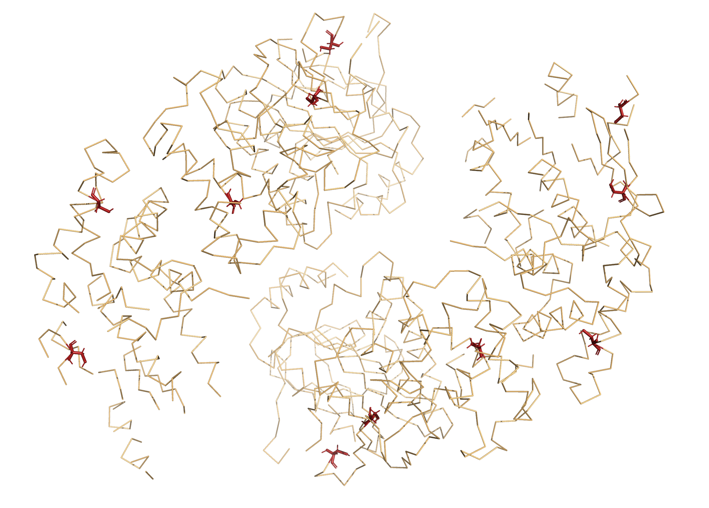
There are two protein chains in one biologcial assembly (heterodimer, fig. 2) that are quite identical in primary structure, but different in secondary and tertiary. Also there are two similar in primary structure biologcial assemblies in one assymetric unit (fig. 1). It has four chains: A and D (fig. 3), B and C in second (fig. 4).
.png)
.png)
.png)
.png)
Chains
The protein is isolated from Thermoanaerobacter pseudethanolicus, but in the article it had also obtained from Escherichia coli by modifying it with the plasmid.
Thermoanaerobacter pseudethanolicus is a thermophilic and strictly anaerobic bacteria that was first isolated from Yellowstone National Park in the United States.
- uniprot_ID: B0KAW2
- Ptotein name: Type IV secretory pathway VirB4 components-like protein.
- Molecular functions: metal ion and nucleotide binding.
Each chain consists of 392 amino acids. There is one modified residue: MSE (selenomethionine, link to .pdb). There are also no mutations in the relative sequence reference in UniProt .
- Chain A: 20 α-helixes and 11 β-sheets (fig. 5A)
- Chain B: 18 α-helixes and 11 β-sheets (fig. 5B)
- Chain C: 16 α-helixes and 6 β-sheets (fig. 5C)
- Chain D: 9 α-helixes and 3 β-sheets (fig. 5D)
.png)
Small molecules
There are water molecules and SO42- ions in 3D structure from PDB (fig.6) but according to article and UniProt the domain can also bind ADP and Mg2+ ions.
- SO4 — SULFATE ION (link to .pdb)
- HOH — Water (link to .pdb)
- GTP — GUANOSINE-5'-TRIPHOSPHATE
- MG — MAGNESIUM ION
.png)
Interactions between aminoacids
Structure of this protein has obtained at pH = 5.75 and all protein interactions will be described for this acidity.
There are such types of protein-protein interactions as hydrogen bonds (including interactions between oppositely charged amino acids) and π-stacking. Disulfide bridges weren't found yet.
There are three types of hydrogen bonds: between backbones (fig. 7 for β-sheets and fig. 8), side chains sensu lato (fig. 9) and oppositely charged side chains (like positive-charged lysine and arginine and negative-charged aspartic and glutamic acids; fig. 10).
.png)
.png)
.png)
.png)
Also we can see π-stacking between amino acids with the aromatic rings in this proteins (like phenylalanine, tyrosine and tryptophan; fig. 11-12). But disulfide bridges aren't in the sctructure: there are only single cysteins in this proteins (fig. 13).
.png)
.png)
References
VirB4 page in Protein Data Bank (PDB, ID: 4AG6) and in UniProt (ID: B0KAW2) .
Walldén, K. et al. (2012). Structure of the VirB4 ATPase, alone and bound to the core complex of a type IV secretion system. PNAS, 109(28), 11348-11353.
Links to .pdb files of selenomethionine, sulfate ion and water.