Algorithms for multiple alignment. Pfam
Last updated: 23-05-2017.
Files to download
Task 1. Multiple alignment algorithms comparison
Homologous proteins from HSP70 family from pr10 were taken for this task. Comparison of alignments by Tcoffee, Muscle and Mafft is presented in Figure 1. It is easy to see that alignments are almost the same, especially if it is a question of domain areas. Most of the differences are located in the end of sequences. For example, comparing Tcoffee and Muscle alignments, there are some differences: Tcoffee row GLY(480), LYS(482), GLY(480), GLY(485), ARG(553) and ARG(511) is Muscle GLY(480), LYS(482), GLY(480), GLY(485), GLY(552) and GLY(510); Tcoffee row ASP(554), ASP(556), ASP(559), ASP(559), ASP(629) and GLN(587) is Muscle ASP(554), ASP(556), ASP(559), ASP(559), GLU(632) and GLU(590); Tcoffee row GLY(480), LYS(482), GLY(480), GLY(485), ARG(553) and ARG(511) is Muscle GLY(480), LYS(482), GLY(480), GLY(485), GLY(552) and GLY(510).

Task 2. Examples of domain architectures
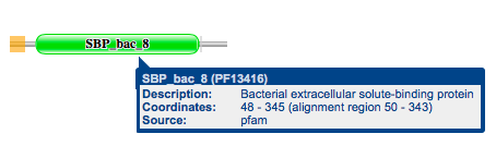
Maltose-binding periplasmic protein (AC P19576) сonsists of a single domain called 'Bacterial extracellular solute-binding protein' (Sample protein Uniprot AC is PF13416) (Fig. 2) [0]. Observed domain is contained in 48 architectures and occurs in 19953 sequences. Detailed information on some of the architectures and domains contained in them is presented in Table 1.
| Sample protein Uniprot AC | Picture of domain architecture | Amount of sequences | Information about domains |
|---|---|---|---|
| A0A0D2U867 | Fig. 3 | 1 | Pkinase_Tyr: a tyrosine kinase is an enzyme that can transfer a phosphate group from ATP to a protein in a cell. It functions as an "on" or "off" switch in many cellular functions. Tyrosine kinases are a subclass of protein kinase [1]. Guanylate_cyc: this domain is responsible for the сatalysis of the cyclization reaction of Adenylate and Guanylate[2] |
| A7BU64 | Fig. 4 | 1 | Peripla_BP_6 is Periplasmic binding protein, as follows from its name, it is responsible for binding something in periplasmic space[3]. |
| A0A0G4IJV7 | Fig. 5 | 1 | 7tm_3 is 7 transmembrane receptor, belongs to metabotropic glutamate family. Structurally it is composed of four elements; an N-terminal signal sequence; a large hydrophilic extracellular agonist-binding region containing several conserved cysteine residues which could be involved in disulphide bonds; a shorter region containing seven transmembrane domains; and a C-terminal cytoplasmic domain of variable length[4] |
Table 1. Detailed information on some of the architectures and domains contained in them.


References
[0] Maltose-binding periplasmic protein, Pfam.
[1] Tyrosine kinase, Pfam.
[2] Adenylate and Guanylate cyclase catalytic domain, Pfam.
[3] Periplasmic binding protein, Pfam.
[4] 7 transmembrane receptor, Pfam.