Transmembrane proteins

Transmembrane proteins


OPM database

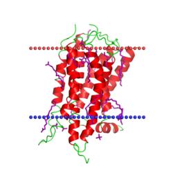
For the task I chose two transmembrane proteins: dopamine D4 receptor[1] and VDAC-1 channel.[2]
  • Hydrophobic Thickness: 31.8 ± 2.0 Å
  • Helix coordinates: A - Tilt: 11° - Segments: 1( 34- 59), 2( 69- 93), 3( 109- 135), 4( 151- 171), 5( 192- 214), 6( 395- 416), 7( 428- 451)
  • Average amount of residues: 22.7
  • Localization: Eukaryotic plasma membrane
  • Species: Homo sapiens
  • Hydrophobic Thickness: 23.4 ± 1.1 Å
  • Helix coordinates:
    X - Tilt: 3° - Segments: 1(26-34), 2(38-47), 3(55-64), 4(69-76), 5(80-88), 6(94-103), 7(111-120),
    8(122-131), 9(137-146), 10(149-156), 11(166-174), 12(178-185), 13(190-197), 14(202-210), 15(218-226), 16(231-238), 17(242-250), 18(255-263), 19(274-282)
  • Average amount of residues: 8.5
  • Localization: Mitochondrial outer membrane
  • Species: Mus musculus

Fig. 1. Dopamine D4 receptor
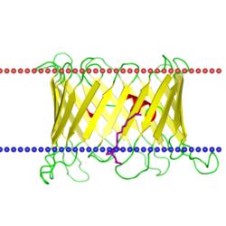
Fig. 2. VDAC-1 channel
TMHMM and Phobius

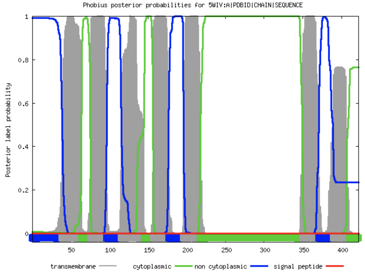
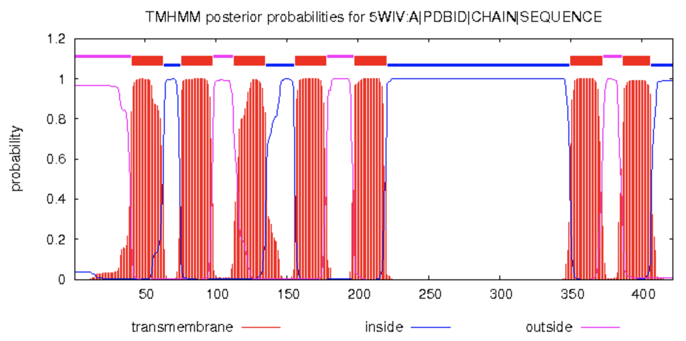
Using 2 services, TMHMM and Phobius, I received 2 images with posterior probabilities for my first transmembrane protein, dopamine D4 receptor.


x-axis - coordinates of aminoacid residues,
y-axis - probability of the transmembrane helix in the place.
Non-cytoplasmic parts marked with blue, cytoplasmic parts - green, signal peptide (i have none) - red, transmembrane - gray.

Fig 3. TMHMM result

Fig 4. Phobius result

x-axis - coordinates of aminoacid residues,
y-axis - probability of the transmembrane helix in the place.
Cytoplasmic parts marked with blue, outside parts - pink, transmembrane - red.

As we can notice, both images are very similar, but the one received from Phobius is more accurate (juste un peu), I think. There were no serious mistakes in results due to the fact that the chosen protein doesn't have a very complex structure.

TCDB

The task was to find chosen proteins in TCDB. I was able to find only the second one, VDAC-1 channel, PDB ID - 3emn.


Pic. 5. TCDB result page

1. - Channels/pores B. - beta-barrel porins 8. - The Mitochondrial and Plastid Porin 1.3 - VDAC1 porin of Bos taurus

Выдача соответствует VDAC1 порину, индентичному человеческому VDAC. У млекопитающих, вообще, 3 известно VDACa (VDAC1, 2 и 3), кодируемые 3 генами, но они очень похожи по последовательности (на ~60-70% идентичны) (Messina et al., 2011). VDAC1 обнаруден квк в мембране митохондрии, так и в клеточной мембране. Возможно, он принимает в участие в формировании рака и болезни Альцгеймера.

References