 Структура белка ацетоацетат декарбоксилазыСтруктура белка ацетоацетат декарбоксилазы
На рис.1 показана структура белка ацетоацетат декарбоксилазы, полученная из файла 3CMB.pdb при помощи программы JMol. С помощью JMol-скрипта атомы хлора покрашены в зелёный цвет, натрия - в сиреневый, α-спирали и β-листы покрашены в разные цвета, β-листы различных цепей отличаются по цвету. Моделью изображения аминокислот, не входящих в состав элементов вторичной структуры, является meshribbon.
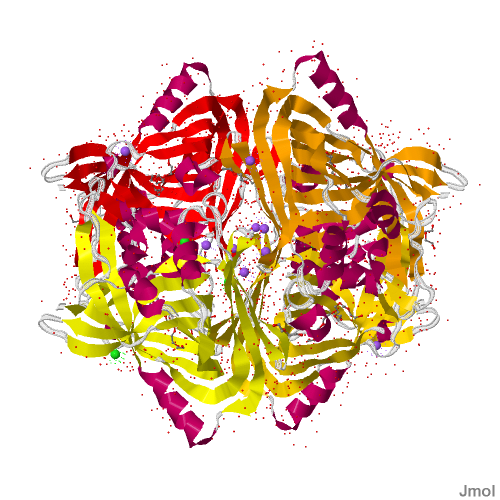
Рисунок 1. Модель белка ацетоацетат декарбоксилазы.
JMol-скрипт, создающий данное изображение, приведён ниже.
load 3CMB.pdb
cpk off
wireframe off
cartoons off
backbone off
select Cl
spacefill IONIC
dots
select Na
spacefill IONIC
dots
select sheet and not :B and not :C and not :D
cartoons
color red
select sheet and not :A and not :C and not :D
cartoons
color orange
select sheet and not :A and not :B and not :D
cartoons
color [255, 215, 0]
select sheet and not :A and not :B and not :C
cartoons
color yellow
select helix
ribbons
color [200, 0, 100]
select protein and not sheet and not helix
meshribbon
color [225, 225, 225]
select HOH
cpk 50
select ligand and not Cl and not Na
cpk 75
wireframe 50
background white
zoom 100
rotate Y 100
rotate X -50
rotate Z 7
Информация о белке, полученная из файла 3CMB.pdb, приведена в таблице 1.
Таблица 1. Информация из файла 3CMB.pdb.
| Title | Crystal structure of acetoacetate decarboxylase (yp_001047042.1) from methanoculleus marisnigri jr1 at 1.60 a resolution |
| Compnd |
Mol_id: 1; molecule: acetoacetate decarboxylase; chain: a, b, c, d; engineered: yes |
| Remark | Resolution. 1.60 angstroms. |
| Hetnam |
	Na sodium ion
        Cl chloride ion
        p33 3,6,9,12,15,18-hexaoxaicosane-1,20-diol
        pe8 3,6,9,12,15,18,21-heptaoxatricosane-1,23-diol
        peg di(hydroxyethyl)ether
|
| Formul |
        mse    28(c5 h11 n o2 se)
        na    12(na 1+)
        cl    4(cl 1-)
        p33    2(c14 h30 o8)
        pe8    2(c16 h34 o9)
        peg    9(c4 h10 o3)
        hoh   *1360(h2 o)
|